03. 2D Medical Images
2D Medical Images
ND320 C2 L1 04 Developing Your Intuition About 2D Medical Images Video
Summary
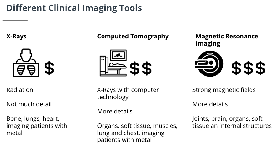
x-ray
This type of imaging projects a type of radiation called x-rays down at the body from a single direction to capture a single image. These are relatively cheap to acquire by imaging standards, but the downside is that they emit radiation and don’t offer too much detail at the organ-level. They allow us to see major structures like bone, lungs, and heart, and they’re safe to use for people who have metal in their bodies.
Computed Tomography (CT)
CT uses x-ray, but they emit x-rays from many different angles around the human body to capture more detail from more different angles. They are more expensive, but they allow us to see more details about organs and soft tissues in the body. Most hospitals in the US have a CT scanner in them.
Magnetic Resonance Imaging (MRI)
MRI uses strong magnetic fields and radio waves to create images of areas of the body from all different angles. It allows us to assess even more details about the human body. This type of imaging is particularly useful for studying the human brain. Although it is safer in that it does not emit radiation, MRI is not safe for people with metal in their bodies. Not all hospitals in the US have MRI scanners and it is a very expensive imaging tool.
Ultrasound
Ultrasound isn't covered in the video. It utilizes high-frequency sound waves beyond the audible limit of human hearing to generate images. Ultrasound waves travel through soft tissues or fluids and bounce back when it hits dense tissues. More waves bounce back if the tissue is denser. The waves that bounce back are captured to generate images. Ultrasound is very safe and commonly used during pregnancy.
2D vs. 3D
Out of these different imaging tools, x-ray is the only 2D imaging tool.
2D imaging takes the picture from a single angle, and everything that is visible to the device at that angle will appear in the picture. You do not see overlapping structures in the 2D image.
3D imaging takes lots of pictures from lots of angles to create a volume of images. You can see structures that are behind one another. The final 3D image is actually a set of 2D images that represent different slices through the body. So, in any single slice or 2D image, you can’t see the whole part but if you scroll through the slices, you will view the volume of that body part.
SOLUTION:
NoNew terms
- X-ray: a 2D imaging technique that projects a type of radiation called x-rays down at the body from a single direction to capture a single image.
- Ultrasound: a 2D imaging technique that uses high-frequency sound waves to generate images.
- Computed Tomography (CT): a 3D imaging technique that emits x-rays from many different angles around the human body to capture more detail from more different angles.
- Magnetic Resonance Imaging (MRI): a 3D imaging technique that uses strong magnetic fields and radio waves to create images of areas of the body from all different angles.
- 2D imaging: an imaging technique that pictures are taken from a single angle.
- 3D imaging: an imaging technique that pictures are taken from different angles to create a volume of images.